Tonic Tensor Tympani Syndrome (TTTS)
Acoustic Reflex and the Stapedius Muscle
Non-acoustic Contraction of Middle Ear Muscles
Tonic Tensor Tympani Syndrome (TTTS)

The following symptoms can occur at the onset of hyperacusis.
- Aural Fullness (Closed Eustachian Tube, contracted tensor tympani, or inner ear pressure)
- Autophony: Boomy or echoey sound from voice (Open Eustachian Tube or conductive hyperacusis)
- Alterations in ventilation of middle ear cavity (Open Eustachian Tube)
- Muffled and/or distorted hearing (When hearing tests are normal)
- Tympanic flutter
- Sharp, Dull, or Burning Pain (Hypothetical Direct and Indirect Mechanisms)
Aural fullness occurs in the majority of cases and is one of the more uncomfortable symptoms that arises when hyperacusis develops. The more severe the hyperacusis the more likely other symptoms from this list will develop. Some researchers refer to this set of symptoms as Tonic Tensor Tympani Syndrome (TTTS). These researchers suspect that TTTS symptoms develop due to unusual hyperactivity in signals sent from the brain to the middle ear muscles to protect the ears from sound. The audiologist Myriam Westcott has led several publications on Acoustic Shock Disorder (ASD) and development of TTTS in hyperacusis and tinnitus patients. These studies provide an interesting perspective on several hyperacusis symptoms (referred to as TTTS symptoms in these studies) that are shared by others who do not have hyperacusis. These researchers suspect that a reduction in anxiety and the desire to protect one’s ears from sound can reduce TTTS symptoms.
The studies by Westcott et al. associate all ear pain in hyperacusis patients to TTTS. This conclusion is inferred from the coinciding TTTS symptoms such as aural fullness and the prevalence of similar ear pain descriptions from non-hyperacusis patients with TTTS. Other researchers, however, disagree with this assessment and do not suspect that pain hyperacusis is a direct product of TTTS. Nonetheless, it is important for those with hyperacusis to recognize the full collection of TTTS symptoms and the role that anxiety and sound tolerance may have in affecting them.
The Tensor Tympani Muscle
The figure at the top of the page shows the location of the tensor tympani muscle in relation to the middle-ear. This muscle will normally tighten the ear drum in specific situations. These include,
- Swallowing
- Speaking, Coughing, Laughing
- Startle Reflex
- Anticipation of loud sounds
 Those with classic TTTS experience spasms or prolonged contraction in this muscle at times other than those listed above. The spasms can be observed by looking at the eardrum through the ear canal using an otoscope and can also be measured using a middle ear impedance test. An example of fluctuating ear impedance caused by TTTS is shown to the right (Klochoff 1979).
“There are individuals in whom the acoustic impedance of the middle ear is not constant in a prolonged recording, which is ordinarily the case, but fluctuates appreciably, quite irregularly and rather slowly. It was concluded that such a very typical impedance fluctuation is caused by spontaneous tonic tensor tympani muscle activity and it was therefore called “THE TONIC TENSOR PHENOMENON”.
Klochoff in Impedance Fluctuation and a “Tensor Tympani Syndrome”
According to the Westcott study, these contractions can lead to the pain felt by many with hyperacusis. They suggest a mechanism similar to that which causes ear pain in TMD might apply to TTTS. There is a striking similarity between some TMD symptoms and TTTS symptoms.
| TTTS | TMD |
| Aural Fullness | Aural Fullness |
| Ear Pain | Ear Pain |
| Muffled Hearing | Muffled Hearing |
| Tinnitus | Tinnitus |
| “Disordered” Balance | Dizziness |
| Pain in Jaw | Pain in Jaw |
| Pain in Neck | Pain in Neck |
| Hyperactive Tensor Tympani | Jaw Clicking, Pain, Stiffness |
Similar to what occurs from TMD, those diagnosed with TTTS can have a radiation of pain or burning sensations in areas innervated by the trigeminal nerve such as the jaw or cheek.
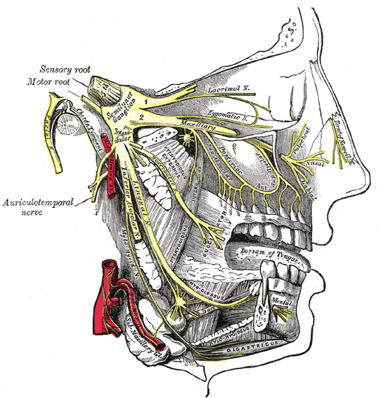
The Trigeminal Nerve
The study suggests that the trigeminal nerve might be irritated by the unusual activity of the tensor tympani which causes referred pain to these areas. The tensor tympani is innervated by the trigeminal nerve as opposed to the stapedius muscle which is innervated by the facial nerve.
Symptom Prevalence
Looking at the data directly can help illustrate just how widespread coinciding TTTS symptoms are in hyperacusis patients. The following plots show the prevalence of TTTS symptoms in the 375 patients surveyed in the 2013 Westcott et. al multi-clinic prevalence study,


Figure from Tonic tensor tympani syndrome in tinnitus and hyperacusis patients by Westcott et al.
The data show that aural fullness and dull ear ache are common symptoms and it also shows that the majority of hyperacusis patients show more than one TTTS symptom. Clearly TTTS symptoms play a part in the experiences of hyperacusis patients. However, whether the hyperactive tensor tympani muscle plays a fundamental role to all symptoms is uncertain. The Westcott study states the following about the mechanisms of TTTS,
“[The hyperactive tensor tympani] appears to initiate physiological reactions in and around the ear without objectively measurable dysfunction or pathology.”
The multi-clinic prevalence study by Westcott et. al can be found here.
In-Depth: Relation to Eustachian Tube Dysfunction, Anxiety, Comments
Tensor Tympani and Eustachian Tube Dysfunction
Role of Anxiety
Comments on TTTS
Tensor Tympani and Eustachian Tube Dysfunction
Several TTTS symptoms such as aural fullness and changes in ventilation are related to the eustachian tube. The eustachian tube is a passageway that connects the upper part of the throat to the middle ear cavity. This tube normally remains closed to prevent bacteria from the throat from traveling to the middle ear, reduce pressure fluctuations, and reduce loudness from self generated sounds. This tube occasionally opens to equalize pressure changes and prevent fluid build up. It will open when you sneeze, swallow, or yawn. If this tube does not open, pressure builds in the middle ear and sensations of aural fullness develop. How can hyperactivity of the tensor tympani have an impact on the eustachian tube?
The opening and closing of the eustachian tube is primarily governed by a portion of the Tensor Veli Palatini (TVP) muscle called the Dilator Tubae. The TVP is connected to the tensor tympani (TT) by a tendon and both muscles often act as a functional unit (Kierner 2002). When you swallow, both the TVP and TT contract, pulling the eardrum tight and opening the eustachian tube. When the TT becomes hyperactive, it is possible that the TVP is impacted and the eustachian tube can be left closed at times when it would normally be open and visa versa. The other end of the TVP is anchored in the soft palate (the soft tissue in the back of the roof of the mouth).
An interactive figure of this eustachian tube dilation can be found here.
More detail on the function of the eustachian tube can be found here.
Anxiety and TTTS
Anxiety and emotional state can play a role in TTTS. Both middle-ear muscles receive direct control from centers of the brain that are influenced by emotional state (the serotonergic system) (McFerran 2007). This along with clinical experience leads some researchers to report that TTTS symptoms can be reduced or eliminated once sound tolerances improve and anxiety to protect one’s ears is reduced.
“Evidence supporting this hypothesis [of anxiety influencing TTTS symptoms] in acoustic shock is seen in the observation that if one person in a call centre suffers an acoustic shock, there is increased risk of other operatives within that centre also developing symptoms. This process could also help explain the patchy distribution of the condition; it is relatively common in some workplaces and geographical locations and completely absent in others”
-Acoustic Shock by McFerran, Baguley
Whether or not removal of anxiety will eliminate TTTS symptoms completely will depend on the particular case. These muscles do not contract solely based on emotional state:
“[In addition to inputs from the cochlear nucleus,] a range of inputs from the superior olivary complex, serotoninergic sources and higher brain centers are thought to descend on TTMNs. The variety of inputs may account for the multifunctional roles of this muscle in response to auditory and non-auditory stimuli.”
-Auditory Brainstem Circuites that mediate the Middle Ear Muscle Reflex by Mukerji et al.
This suggests that anxiety can significantly influence TTTS symptoms but that it cannot be used as a blanket explanation.
Comments on TTTS
Hyperactivity of the tensor tympani is not a requirement for TTTS diagnosis in these studies. Rather, a patient is diagnosed with TTTS if one or more unexplained otological symptoms are present. While symptoms such as tympanic flutter can clearly be attributed to the tensor tympani muscle, others such as aural fullness or ear pain can have multiple causes. Thus it seems possible that, in these studies, TTTS may be a catch-all diagnosis for unexplained otological symptoms without means of identifying a true root cause. The limited amount of literature on TTTS suggests this diagnosis is not commonly used.
The tensor tympani is not a significant part of the acoustic reflex which is why the recent hyperacusis literature review by Tyler et al noted the logic was unclear for how the tensor tympani could be responsible for sound-induced pain. Many with hyperacusis feel pain without feeling anxious about sound. Many do not feel pain when anticipating loud sounds. It is also not common for those with hyperacusis to feel pain while swallowing, which will cause the tensor tympani to contract. This suggests that if sound-induced pain is triggered by middle ear muscles, it is only triggered by unusually strong contractions or change in equilibrium. Such contractions should be measurable. Applying indirect TTTS pain mechanisms such as irritation of the trigeminal nerve to pain hyperacusis still requires an explanation for the pathway from sound to the tensor tympani when anxiety and startle are absent. Some researchers suspect that unusual activation of the stapedius muscle, while not formally categorized as part of TTTS, may contribute to sound-induced pain. More details of the role of the stapedius in sound induced pain along with counterarguments are presented in the next section.
The 2013 multi-clinic study by Westcott et. al. found that 81% of hyperacusis patients had TTTS. They determined this not by examining for hyperactivity of the tensor tympani but by seeing if one or more unexplained TTTS symptoms was present. Thus, almost any hyperacusis patient with unexplained ear pain would be diagnosed with TTTS. As the fundamental characteristic of those with pain hyperacusis is ear pain, one must be careful to lump this as a side effect of TTTS. It is likely that a hyperactive tensor tympani can cause ear pain but there is a possibility that ear pain independent of TTTS is getting included in this study.
There are claims from those who focus on TTTS research that pain hyperacusis is not just influenced by anxiety but is actually caused by anxiety. Supporting evidence for this claim was not presented. Many with hyperacusis make clear that anxiety develops after tolerance levels have already dropped.
Nonetheless, the prevalence of coinciding TTTS symptoms in hyperacusis patients is important. Those who have developed hyperacusis may find some comfort in having several of their symptoms described as a product of TTTS.
Acoustic Reflex and the Stapedius Muscle

There are two muscles in the middle ear that help regulate the sound that is sent to the inner ear. The first is the tensor tympani that was discussed in the previous section. The second is the body’s smallest muscle; the stapedius. In humans, the stapedius muscle reacts to sound as part of the acoustic reflex (which is also referred to as the stapedius reflex). When the stapedius contracts, it pulls the stapes bone away from the cochlea which attenuates mostly low frequencies. The tensor tympani, on the other hand, shows only a weak response to sound. The function of the stapedius is to protect the ear from loud sounds and to help filter for speech by filtering low frequency sounds. The stapedius will start to contract at around 85 dB and will gradually increase its pull as sound intensity increases.
The acoustic reflex could contribute to hyperacusis in one of two ways. First, if the acoustic reflex is not functioning (for example if the stapedius tendon is cut), then loud sounds will not be attenuated as they were before. This can initiate a degree of loudness hyperacusis. The second way the acoustic reflex might impact hyperacusis is if the contraction of the stapedius muscle somehow triggered pain. If this were to occur, then sounds above the acoustic reflex threshold would be sensed as damaging. This second scenario, of a pain-inducing acoustic reflex, is not suspected to be a cause of pain hyperacusis in the literature. Such a cause would have tell-tale signs. As the stapedius contracts during speech, pain while speaking may be expected. A pain-inducing acoustic reflex would also trigger pain in both ears even if the sound was sent to only one ear. As those with hyperacusis generally show normal or high acoustic reflex thresholds, only high intensity sounds would trigger contraction.
In-Depth: Stapedius Innervation, Acoustic Reflex
Innervation of the Stapedius Muscle
The stapedius is innervated by a branch of the facial nerve (Cranial nerve VII). Pain receptors have not been found in the stapedius muscle. Pain receptors would be a bad idea given the potential for pain hyperacusis and the fact that these muscles are involuntary. Rats, for example, have no sensory nerves at all in their middle ear muscles. In humans, the stapedius is overwhelmingly innervated by motor neurons but it does have a small collection of sensory neurons (Belvins 1967). These are muscle spindles which detect muscle length. The function of the “thin, filamentous nerve fibers” found in the stapedius tendon is unknown.
The Acoustic Reflex
The path of the acoustic reflex is relatively short. Sound travels through the middle ear to the cochlea, then travels through the auditory nerve to brainstem (the ventral cochlear nucleus->superior olive->facial nerve neurons), and then through the facial nerve to the stapedius muscle.

While this path is short, studies from Munro and Blount have shown that there is potential for gain adaptation through use of earplugs or sound generators. Average acoustic reflex threshold changes of 5 dB were observed within one week of earplug use.
 In normal ears, the acoustic reflex can vary from 70dB to 90 dB using a single tone. Similar to LDLs, acoustic reflex thresholds are similar across frequency. If wider bandwidth signals such as speech or broadband noise are used, acoustic reflex thresholds drop to a range of roughly 70dB-75dB. After sound passes this threshold, the stapedius will gradually increase its pull as sound intensity increases. The plot to the right (Sprague 1981) is an example of acoustic impedance change caused by the stapedius increasing its pull with increasing sound intensity. It has been suggested that some symptoms of hyperacusis patients may be the result of an increase in slope and peak magnitude of this plot (impedance vs sound level). Measurements to support this theory have not been published.
Acoustic Reflexes and Loudness Discomfort Levels
There are conflicting messages in the literature regarding how well acoustic reflexes thresholds (ARTs) correlate to loudness discomfort levels (LDLs) of normal ears. Olsen performed a meta analysis on several studies and concluded that for those with normal hearing, the LDLs and ARTs measured were uncorrelated. Other studies have found that a correlation can be found. Regardless of the relationship between LDLs and ARTs in normal hearing individuals, the metrics need to be compared among those who have been diagnosed with hyperacusis. A 1999 study led by Anari measured ARTs of 51 hyperacusis patients (the remaining 49 patients did not undergo this loud test). All ARTs were found to be within normal limits (between 70dB and 95dB). Correlation measures were not reported.
Interestingly, a 2020 study by Saxena found that those with hyperacusis had slightly higher ARTs when compared to those without hyperacusis.

Data interpreted from figure 1 from Saxena et. al.
The Saxena study also found that the reflex amplitude was weaker on average in those with hyperacusis. The above study defined hyperacusis as both a clinical diagnosis of hyperacusis and a Hyperacusis Questionnaire (HQ) score > 28. The results above complicate the central gain theory of hyperacusis. Overprotection studies by Munro found that acoustic reflex thresholds decreased in those with normal hearing who overused earplugs for 7 days until they were removed for ART testing. The Munro study concluded there was thus a gain increase in the brainstem to compensate for the earplug usage. The Saxena study shows the opposite trend in hyperacusis patients. It is possible that this discrepancy was due to attrition bias (dropouts) in the Saxena study; a result that would be interesting in its own right.
Ipsilateral vs Contralateral Acoustic Reflex
There are two types of acoustic reflexes. There is the ipsilateral acoustic reflex and the contralateral acoustic reflex. The ipsilateral reflex is the stapedius contraction on the same ear that is receiving sound. The contralateral reflex is the stapedius contraction on the ear opposite of that which is receiving sound. Contralateral reflexes and ipsilateral reflexes trigger at loudness levels around 85 dB with contralateral thresholds usually 3 dB higher than ipsilateral thresholds. As a result, if hearing protection is worn in only one ear, sound from the unprotected ear would still be able to trigger the stapedius reflex in the protected ear.
Non-acoustic Contraction of Middle Ear Muscles
The middle ear muscles do not require sound to contract. Swallowing will contract the tensor tympani and tensor veil palatini. The stapedius and tensor tympani will contract as a result of the palpebral reflex or if a puff of air is sent to the eyes with a bulb syringe (Fee 1981).
Have ideas on how to make this article better? Please contact improve@hyperacusisfocus.org.
References
Anari M, Axelsson Alf, Eliasson A, Magnusson L. Hypersensitivity to Sound: Questionnaire data, audiometry and classification. Scand Audiol 1999:28:219-230
Al-azazi M, Othman B. Acoustic reflex threshold and loudness discomfort. Saudi Medical Journal 2000:21:251-256.
Arrestguieta L, Acuna L, Ortiz G. Tensor veli palatini and tensor tympani muscles: Anatomical, functional and symptomatic links. Acta Otorrinolaringologica Espanola 2010:61:26-33.
Belvins C, Innervation patterns of the human stapedius muscle. Arch Otolaryng 1967:86:136-142.
Borg E, Counter S. The Middle-Ear Muscles. Scientific American 1989:August:74-80.
Fee W. Clinical Application of Nonacoustic Middle Ear Muscle Stimulation. Arch Otolaryngol 1981:107:224-226. 1981:107:224-226.
Hain T, http://dizziness-and-balance.com/disorders/hearing/tinnitus/tensor%20tympani%20and%20stapedius%20myoclonus%20tinnitus.html
Hain T, http://dizziness-and-balance.com/testing/acoustic_reflexes.html
Kierner AC, Mayer R, Kirschhofer K. Do the tensor tympani and tensor veli palatini muscles of man form a functional unit? A histochemical investigation of their putative connections. Hearing Research 2002:165:48-52.
Klochoff I. Impedance fluctuation and a “Tensor Tympani Syndrome” Proceedings of the 4th International Symposium on Acoustic Measurements. Lisbon. 1979:69-76.
McFerran DJ, Baguley DM. Acoustic Shock. The Journal of Laryngology & Otology 2007:121:301-305.
Mukerji S, Windsor A, Lee D. Auditory Brainstem Circuits That Mediate the Middle Ear Muscle Reflex. Trends in Amplification 2010:14(3):170-191.
Munro K, Blount J. Adaptive plasticity in brainstem of adult listeners following earplug-induced deprivation. J. Acoust. Soc. Am. 2009:126:568-571.
Munro K, Blount J. Brainstem plasticity and modified loudness following short-term use of hearing-aids. J. Acoust. Soc. Am. 2013:133:343.
Olsen, S. The relationship between the uncomfortable loudness level and the acoustic reflex threshold for pure tones in normally-hearing and impaired listeners – A meta analysis. Audiology 1999:38:61-68.
Ramirez et. al. Topical review: Temporomandibular disorders in an integral otic system model. International Journal of Audiology 2008:47:215-227.
Saxena U, Singh B, Kumar S.B., Chacko G, Bharath K.N.S.V. Acoustic Reflexes in Individuals Having Hyperacusis of the Auditory Origin. Indian J Otolaryngol Head Neck Surg 2020:72:497-502.
Sprague B, Wiley T, Block M. Dynamics of Acoustic Reflex Growth. Audiology 1981:20:15-40.
Tyler R, Pienkowski M, et. al. A Review of Hyperacusis and Future Directions: Part I. Definitions and Manifestations. American Journal of Audiology 2014:23:402-419.
Tyler R, Pienkowski M, et. al. A Review of Hyperacusis and Future Directions: Part II. Measurement, Mechanisms, and Treatment. American Journal of Audiology 2014:23:420-436.
Westcott M et. al. Tonic tensor tympani syndrome in tinnitus and hyperacusis patients: A multi-clinic prevalence study. Noise & Health 2013:15:117-128.
Westcott M. Acoustic shock disorder. Tinnitus discovery-Asia and pacific tinnitus symposium Auckland 2010:123:25-31.